Artroscopia Pediatrica

Artroscopia Pediatrica

lunes, 24 de febrero de 2014

Arthroscopic debridement in the treatment of patients with osteoarthritis of the elbow, based on computer simulation

http://www.bjj.boneandjoint.org.uk/content/96-B/2/237.abstract


Arthroscopic debridement in the treatment of patients with osteoarthritis of the elbow, based on computer simulation

  1. K. Shimada, MD, PhD, Orthopaedic Surgeon, Division Chief2;
  2. K. Oka, MD, PhD, Orthopaedic Surgeon, Assistant Professor1;
  3. H. Tanaka, MD, PhD, Orthopaedic Surgeon, Assistant Professor1;
  4. K. Sugamoto, MD, PhD, Orthopaedic Surgeon, Professor3;
  5. H. Yoshikawa, MD, PhD, Orthopaedic Surgeon, Professor1; and
  6. T. Murase, MD, PhD, Orthopaedic Surgeon, Associate Professor1
+ Author Affiliations
  1. 1Osaka University Graduate School of Medicine, Department of Orthopaedic Surgery, 2-2 Yamadaoka, Suita, Osaka 565-0871, Japan.
  2. 2Osaka Koseinenkin Hospital, Department of Orthopaedic Surgery, 4-2-78 Fukushima, Osaka 553-0003, Japan.
  3. 3Osaka University Graduate School of Medicine, Department of Orthopaedic Biomaterial Science, 2-2 Yamadaoka, Suita, Osaka 565-0871, Japan.
  1. Correspondence should be sent to Dr J. Miyake; e-mail: miyake-osk@umin.ac.jp

Abstract

We retrospectively assessed the value of identifying impinging osteophytes using dynamic computer simulation of CT scans of the elbow in assisting their arthroscopic removal in patients with osteoarthritis of the elbow. A total of 20 patients were treated (19 men and one woman, mean age 38 years (19 to 55)) and followed for a mean of 25 months (24 to 29). We located the impinging osteophytes dynamically using computerised three-dimensional models of the elbow based on CT data in three positions of flexion of the elbow. These were then removed arthroscopically and a capsular release was performed.
The mean loss of extension improved from 23° (10° to 45°) pre-operatively to 9° (0° to 25°) post-operatively, and the mean flexion improved from 121° (80° to 140°) pre-operatively to 130° (110° to 145°) post-operatively. The mean Mayo Elbow Performance Score improved from 62 (30 to 85) to 95 (70 to 100) post-operatively. All patients had pain in the elbow pre-operatively which disappeared or decreased post-operatively. According to their Mayo scores, 14 patients had an excellent clinical outcome and six a good outcome; 15 were very satisfied and five were satisfied with their post-operative outcome.
We recommend this technique in the surgical management of patients with osteoarthritis of the elbow.
Cite this article: Bone Joint J 2014;96-B:237–41.

Footnotes

  • We thank Ryoji Nakao, computer programmer; Sumika Ikemoto, clinical assistant at the Department of Orthopedic Surgery, Osaka University Graduate School of Medicine; Hisao Moritomo, MD, PhD at the Department of Physical Therapy, Osaka Yukioka College of Health Science; Nobuyuki Kubo, MD at the Department of Orthopedic Surgery, Yukioka Hospital; Eiji Sogo, MD at the Department of Orthopedic Surgery, Kita-Osaka Police Hospital, for their contributions to this study.
    No benefits in any form have been received or will be received from a commercial party related directly or indirectly to the subject of this article.
    This article was primary edited by P. Baird and first-proof edited by J. Scott.
  • Received August 18, 2012.
  • Accepted October 22, 2013.

Controlar el dolor de la cirugía de pie y tobillo / Controlling Pain from Foot and Ankle Surgery

http://icjr.net/perioppearl_3_calcaneal_osteotomy.htm


Controlling Pain from Foot and Ankle Surgery

Controlar el dolor de la cirugía de pie y tobillo
Selene G. Parekh , MD , MBA , de la Duke University Medical Center en Durham, Carolina del Norte, ha comenzado a utilizar la infiltración local con liposomas bupivacaína suspensión inyectable ( EXPAREL ®) , un anestésico local que tiene propiedades analgésicas para hasta 72 horas después de la administración , para proporcionar el control del dolor de sus pacientes cirugía de pie y tobillo.
En un video recientemente publicado en el Centro de Dolor de Recursos para la administración Periop , Dr. Parekh demuestra su técnica de infiltración local con un paciente que se somete a una osteotomía del calcáneo . Para la infiltración , el Dr. Parekh utiliza dos jeringas de 10 ml de la solución de bupivacaína liposómica con agujas de calibre 25 . El tejido blando se derrumba alrededor de una aguja de calibre 25 , dijo, lo que evitará que la solución se filtre fuera del tejido .
Usando una técnica de aguja que se mueve , el Dr. Parekh inyecta en el tejido subcutáneo alrededor de la herida quirúrgica para infiltrarse en las ramas del nervio peroneo , ramas nerviosas safena y ramas del nervio tibial . Se inserta la aguja en el cubo y luego inyecta la solución mientras se tira de la aguja .
Una vez que la incisión se ha cerrado , se inyecta el resto de la solución paralela a la incisión para cubrir el nervio sural .
Selene G. Parekh, MD, MBA, from Duke University Medical Center in Durham, North Carolina, has begun using local infiltration with bupivacaine liposome injectable suspension (EXPAREL®), a local anesthetic that has analgesic properties for up to 72 hours following administration, to provide pain control for his foot and ankle surgery patients.
In a video recently posted on the Periop Pain Management Resources Center, Dr. Parekh demonstrates his local infiltration technique with a patient who is undergoing a calcaneal osteotomy. For the infiltration, Dr. Parekh uses two 10-mL syringes of the liposomal bupivacaine solution with 25-gauge needles. The soft tissue will collapse around a 25-gauge needle, he said, which will prevent the solution from leaking out of the tissue.
Using a moving needle technique, Dr. Parekh injects into the subcutaneous tissue around the surgical wound to infiltrate the peroneal nerve branches, saphenous nerve branches, and tibial nerve branches. He inserts the needle to the hub and then injects the solution as he pulls the needle out.
Once the incision has been closed, he injects the remainder of the solution parallel to the incision to cover the sural nerve.

sábado, 22 de febrero de 2014

Esguinces de tobillo


Las tasas de tratamiento de la osteoporosis después de una fractura de cadera en declive / Rates of osteoporosis treatment after hip fracture on the decline

http://www.healio.com/orthopedics/arthritis/news/online/%7B93c433c9-d99f-480c-a93c-cecfa6180f9f%7D/rates-of-osteoporosis-treatment-after-hip-fracture-on-the-decline


Las tasas de tratamiento de la osteoporosis después de una fractura de cadera en declive


La mayoría de los pacientes con fractura de cadera no recibe tratamiento para la osteoporosis y las tasas de tratamiento han disminuido , un hallazgo que no es consistente con las guías de tratamiento de la osteoporosis avalados por la Fundación Nacional de Osteoporosis , según un estudio publicado recientemente .

"La mayoría de los pacientes que sufren una fractura de cadera no usan medicamento para la osteoporosis en el año siguiente y los tipos de tratamiento han empeorado , Daniel H. Solomon , MD, MPH, del Hospital Brigham y de Mujeres , y sus colegas , declaró en abstracto.

Los investigadores llevaron a cabo un estudio observacional de cohorte retrospectivo basado en los Estados Unidos los datos de reclamaciones de seguros administrativa para los beneficiarios con comercial de seguro de salud suplementario de Medicare . El estudio incluyó a 96.887 participantes que fueron hospitalizados por una fractura de cadera entre 1 de enero 2002 y el 31 de diciembre de 2011, y de 50 años o de más edad en la admisión. Resultado de interés incluyen el uso de medicamentos osteoporosis dentro de los 12 meses después del alta . Investigadores censurados los pacientes después de 12 meses , la pérdida para el seguimiento o un reclamo médico para el cáncer o la enfermedad de Paget , lo que ocurra primero evento .

En general , el 35,5 % de los pacientes fueron censurados antes de llegar a los 12 meses de seguimiento. Los investigadores encontraron un 28.5 % de probabilidad de medicamento para la osteoporosis utilizar dentro de los 12 meses después del alta según las estimaciones de Kaplan -Meier. Las tasas disminuyeron de 40.2 % en 2002 a 20,5% en 2011. Características de los pacientes , incluyendo la edad avanzada y el sexo masculino , se asociaron con una menor probabilidad de utilización de los medicamentos osteoporosis. Sin embargo , los investigadores encontraron el uso de medicamentos de la osteoporosis antes de la fractura era más fuerte y más directamente asociado con el uso de medicación de la osteoporosis después de la fractura .

Rates of osteoporosis treatment after hip fracture on the decline


The majority of patients with hip fracture do not receive osteoporosis treatment and the rates of treatment have been declining, a finding that is not consistent with osteoporosis treatment guidelines endorsed by the National Osteoporosis Foundation, according to a recently published study.
“Most patients suffering a hip fracture do not use osteoporosis medication in the subsequent year and treatment rates have worsened, Daniel H. Solomon, MD, MPH, from Brigham and Women's Hospital, and colleagues, stated in the abstract.
Researchers conducted a retrospective, observational cohort study based on U.S. administrative insurance claims data for beneficiaries with commercial of Medicare supplemental health insurance. The study included 96,887 participants who were hospitalized for a hip fracture between Jan. 1, 2002, and Dec. 31, 2011, and aged 50 years or older at admission. Outcome of interest included osteoporosis medication use within 12 months after discharge. Researchers censored patients after 12 months, loss to follow-up or a medical claim for cancer or Paget’s disease, whichever event occurred first.
Overall, 35.5% of patients were censored before reaching 12 months of follow-up. Researchers found a 28.5% probability of osteoporosis medication use within 12 months after discharge as estimated by Kaplan-Meier. The rates declined from 40.2% in 2002 to 20.5% in 2011. Patient characteristics, including older age and male gender, were associated with reduced likelihood of osteoporosis medication use. However, researchers found osteoporosis medication use before the fracture was most strongly and most positively associated with the use of osteoporosis medication after fracture.

La fascitis plantar / Plantar fasciitis

http://www.ncbi.nlm.nih.gov/pubmedhealth/PMH0004438/

La fascitis plantar

Última revisión : marzo 1, 2012.

La fascitis plantar es una inflamación del tejido grueso en la parte inferior del pie. Este tejido se denomina fascia plantar. Se conecta el hueso del talón a los dedos y crea el arco del pie .
fascia plantar

Causas , incidencia y factores de riesgo

La fascitis plantar ocurre cuando la banda gruesa de tejido en la parte inferior del pie se estira demasiado o muy usada . Esto puede ser doloroso y hacer más difícil el caminar .

La fascitis plantar

Usted es más propenso a la fascitis plantar si usted tiene:

Problemas con el arco del pie (tanto pie plano y arcos elevados )

Carreras de larga distancia , especialmente correr cuesta abajo o en superficies desiguales

Aumento repentino de peso o la obesidad

Tendón de Aquiles (el tendón que conecta los músculos de la pantorrilla al talón )

Los zapatos con poco arco de soporte o suelas blandas

La fascitis plantar es visto tanto en hombres como en mujeres. Sin embargo, con mayor frecuencia afecta a hombres entre activos 40 - 70 . Es una de las dolencias ortopédicas más comunes relacionadas con el pie .

La fascitis plantar es comúnmente considerado como causada por un espolón en el talón , pero las investigaciones han encontrado que este no es el caso . En las radiografías , espolones del talón se observan en personas con y sin fascitis plantar.

síntomas

La queja más común es el dolor y la rigidez en la parte inferior del talón . El dolor en el talón puede ser sordo o agudo. La parte inferior del pie también puede doler o arder .
El dolor suele ser peor :

Por la mañana, cuando usted toma sus primeros pasos

Después de estar de pie o sentado durante un tiempo

Al subir las escaleras

Después de una intensa actividad

El dolor puede desarrollarse lentamente con el tiempo, o de repente después de una actividad intensa.


Signos y exámenes

El médico llevará a cabo un examen físico . Esto puede mostrar:

Sensibilidad en la parte inferior de su pie

Los pies planos o arcos altos

Hinchazón de los pies leve o enrojecimiento

Rigidez o tensión del arco en la parte inferior de su pie.

Las radiografías pueden ser tomadas para descartar otros problemas.


tratamiento

El médico suele recomendar primero :

El acetaminofeno (Tylenol ) o ibuprofeno ( Advil , Motrin ) para reducir el dolor y la inflamación

Talón y pie ejercicios de estiramiento

Las férulas nocturnas para usar mientras se duerme para estirar el pie

En reposo tanto como sea posible durante al menos una semana

El uso de zapatos con buen soporte y cojines

Otras medidas para aliviar el dolor incluyen:

Aplicar hielo en la zona dolorida. Haga esto por lo menos dos veces al día durante 10 - 15 minutos , más a menudo en el primer par de días.

Trate de usar un soporte para el talón , almohadillas de fieltro en el área del talón , o el calzado.

Utilice férulas nocturnas para estirar la fascia lesionada y permitir que sane .

Si estos tratamientos no funcionan , el médico puede recomendar:


El uso de un elenco de arranque, que luce como una bota de esquí, durante 3-6 semanas . Se puede extraer para el baño.

Dispositivos para el calzado hecho a medida ( plantillas ortopédicas )

Inyecciones de esteroides o inyecciones en el talón

A veces , se necesita cirugía del pie.

Expectativas ( pronóstico)

Los tratamientos no quirúrgicos casi siempre mejoran el dolor. El tratamiento puede durar desde varios meses hasta 2 años antes de que los síntomas mejoren . La mayoría de los pacientes se sienten mejor en 9 meses. Algunas personas necesitan una cirugía para aliviar el dolor.


Complicaciones

El dolor puede continuar a pesar del tratamiento . Algunas personas pueden necesitar cirugía. La cirugía tiene sus propios riesgos. Hable con su médico acerca de los riesgos de la cirugía .
Situaciones que requieren asistencia médica

Póngase en contacto con su proveedor de atención médica si se presentan síntomas de la fascitis plantar.

prevención

Asegurarse de que su tobillo , tendón de Aquiles y los músculos de la pantorrilla son flexibles puede ayudar a prevenir la fascitis plantar.
A.D.A.M. Medical Encyclopedia.

Plantar fasciitis

Last reviewed: March 1, 2012.
Plantar fasciitis is inflammation of the thick tissue on the bottom of the foot. This tissue is called the plantar fascia. It connects the heel bone to the toes and creates the arch of the foot.
Plantar fascia

Causes, incidence, and risk factors

Plantar fasciitis occurs when the thick band of tissue on the bottom of the foot is overstretched or overused. This can be painful and make walking more difficult.
Plantar fasciitis
You are more likely to get plantar fasciitis if you have:
  • Foot arch problems (both flat feet and high arches)
  • Long-distance running, especially running downhill or on uneven surfaces
  • Sudden weight gain or obesity
  • Tight Achilles tendon (the tendon connecting the calf muscles to the heel)
  • Shoes with poor arch support or soft soles
Plantar fasciitis is seen in both men and women. However, it most often affects active men ages 40 - 70. It is one of the most common orthopedic complaints relating to the foot.
Plantar fasciitis is commonly thought of as being caused by a heel spur, but research has found that this is not the case. On x-ray, heel spurs are seen in people with and without plantar fasciitis.

Symptoms

The most common complaint is pain and stiffness in the bottom of the heel. The heel pain may be dull or sharp. The bottom of the foot may also ache or burn.
The pain is usually worse:
  • In the morning when you take your first steps
  • After standing or sitting for a while
  • When climbing stairs
  • After intense activity
The pain may develop slowly over time, or suddenly after intense activity.

Signs and tests

The health care provider will perform a physical exam. This may show:
  • Tenderness on the bottom of your foot
  • Flat feet or high arches
  • Mild foot swelling or redness
  • Stiffness or tightness of the arch in the bottom of your foot.
X-rays may be taken to rule out other problems.

Treatment

Your health care provider will usually first recommend:
  • Acetaminophen (Tylenol) or ibuprofen (Advil, Motrin) to reduce pain and inflammation
  • Heel and foot stretching exercises
  • Night splints to wear while sleeping to stretch the foot
  • Resting as much as possible for at least a week
  • Wearing shoes with good support and cushions
Other steps to relieve pain include:
  • Apply ice to the painful area. Do this at least twice a day for 10 - 15 minutes, more often in the first couple of days.
  • Try wearing a heel cup, felt pads in the heel area, or shoe inserts.
  • Use night splints to stretch the injured fascia and allow it to heal.
If these treatments do not work, your health care provider may recommend:
  • Wearing a boot cast, which looks like a ski boot, for 3-6 weeks. It can be removed for bathing.
  • Custom-made shoe inserts (orthotics)
  • Steroid shots or injections into the heel
Sometimes, foot surgery is needed.

Expectations (prognosis)

Nonsurgical treatments almost always improve the pain. Treatment can last from several months to 2 years before symptoms get better. Most patients feel better in 9 months. Some people need surgery to relieve the pain.

Complications

Pain may continue despite treatment. Some people may need surgery. Surgery has its own risks. Talk to your doctor about the risks of surgery.

Calling your health care provider

Contact your health care provider if you have symptoms of plantar fasciitis.

Prevention

Making sure your ankle, Achilles tendon, and calf muscles are flexible can help prevent plantar fasciitis.

References

  1. Wapner KL, Parekh SG. Heel pain. In: DeLee JC, Drez D Jr, Miller MD, eds. DeLee and Drez’s Orthopaedic Sports Medicine. 3rd ed. Philadelphia, Pa: Saunders Elsevier; 2009:section F.
  2. Abu-Laban RV, Ho K. Ankle and foot. In: Marx JA, Hockberger RS, Walls RM, et al, eds. Rosen’s Emergency Medicine: Concepts and Clinical Practice. 7th ed. Philadelphia, Pa: Mosby Elsevier; 2009:chap 55.
  3. Silverstein JA, Moeller JL, Hutchinson MR.Common issues in orthopedics. In: Rakel RE, ed. Textbook of Family Medicine. 8th ed. Philadelphia, Pa: Saunders Elsevier;2011:chap 30.
Review Date: 3/1/2012.
Reviewed by: Linda J. Vorvick, MD, Medical Director and Director of Didactic Curriculum, MEDEX Northwest Division of Physician Assistant Studies, Department of Family Medicine, UW Medicine, School of Medicine, University of Washington. C. Benjamin Ma, MD, Assistant Professor, Chief, Sports Medicine and Shoulder Service, UCSF Department of Orthopaedic Surgery. Also reviewed by David Zieve, MD, MHA, Medical Director, A.D.A.M., Inc.
Logo of A.D.A.M.
Copyright © 2013, A.D.A.M., Inc.

Recuperación de una artrocentesis / Arthrocentesis Recovery

http://www.arthritis-health.com/treatment/joint-aspiration/arthrocentesis-recovery-and-potential-risks

Arthrocentesis Recovery

Algunos pacientes pueden ser advertidos de que van a necesitar que alguien lo lleve a casa después del procedimiento. Los anestésicos locales como la lidocaína generalmente desaparecen en 2 a 4 horas , por lo que los pacientes pueden sentir un aumento del dolor poco después de salir office.2 del médico Es común tener dolor leve durante un día o dos después del procedimiento .

Los pacientes deben preguntar a sus médicos si está bien tomar medicamentos antiinflamatorios ( AINES ) u otros analgésicos de venta libre . Dependiendo de las circunstancias , el médico puede recomendar la formación de hielo periódicamente la junta y envolverlo en un vendaje para evitar que vuelva a la hinchazón.

Véase Cómo cuidar a una rodilla hinchada Artrocentesis Posibles riesgos y complicaciones. El efecto secundario más común asociado con la artrocentesis es molestia articular temporal. Otros riesgos incluyen :

La aguja podría raspar o perforar los tendones de la articulación, los vasos sanguíneos o los nervios.

El paciente puede tener una reacción alérgica a la anestesia.

En casos raros, el pinchazo de la aguja puede introducir sangre o infección en la cavidad articular thesynovial . La infección se produce en menos de 0,01 % de los pacientes que se someten a injections.3 conjunta

Llame a un médico inmediatamente si presenta alguno de los siguientes:

Dolor moderado a severo ( dolor leve y el dolor es normal , pero el dolor que no se controla

adecuadamente con la medicación over-the- counter del dolor y bolsas de hielo deben ser reportados )

Moderada a extrema hinchazón

La decoloración o erupción alrededor de la zona de la inyección

fiebre

Sangrado o secreción del punto de entrada de la aguja

Para la mayoría de los pacientes , la artrocentesis es un procedimiento seguro y sin incidentes.
Some patients may be advised that they will need someone to drive them home following the procedure. Local anesthetics such as lidocaine typically wear off in 2 to 4 hours, so patients may feel an increase in pain shortly after leaving the doctor’s office.2
It is common to have mild soreness for a day or two after the procedure.
Patients should ask their doctors whether it is okay to take anti-inflammatory medication (NSAIDs) or other over-the-counter pain medication. Depending on the circumstances, the doctor may recommend periodically icing the joint and wrapping it in a bandage to prevent re-swelling.

Arthrocentesis Potential Risks and Complications

The most common side effect associated with arthrocentesis is temporary joint discomfort. Other risks include:
  • The needle may scrape or puncture the joint’s tendons, blood vessels or nerves.
  • The patient may have an allergic reaction to the anesthetic.
  • In rare cases, the needle puncture can introduce blood or infection into thesynovial joint cavity. Infection occurs in less than 0.01% of patients who undergo joint injections.3
Call a doctor immediately if any of the following occur:
  • Moderate to severe pain (mild pain and soreness is normal, but pain that is not adequately controlled with over-the-counter pain medication and ice packs should be reported)
  • Moderate to extreme swelling
  • Discoloration or rash around the injection area
  • Fever
  • Bleeding or discharge from the needle’s entry point
For most patients, arthrocentesis is a safe and uneventful procedure.
References
  1. Courtney P, Doherty M. Joint aspiration and injection and synovial fluid analysis. Best Practice & Research Clinical Rheumatology, Volume 27, Issue 2, April 2013, Pages 137-169, ISSN 1521-6942, www.sciencedirect.com.
  2. Zuber TJ. Knee joint aspiration and injection. Am Fam Physician. 2002 Oct 15;66(8):1497-500, 1503-4, 1507. PubMed PMID: 12408424.

Osteopoikilosis

http://www.learningradiology.com/archives2010/COW%20415-Osteopoikilosis/osteopoikcorrect.htm


Osteopoikilosis
"Spotted Bone Disease"
General Considerations
  • Rare autosomal dominant or sporadic osteosclerotic dysplasia
  • Multiple punctate sclerotic lesions representing "bone islands" or foci of compact bone located in cancellous bone
  • Asymptomatic
  • Occurs in the epiphyses and metaphyses with predilection for
    • Tubular bones of the hands and feet
    • Carpals
    • Tarsals
    • Pelvis
    • Scapula
  • Ribs, clavicles, spine, and skull are rarely involved
  • Usually clustered around joints
  • Males and females affected equally
Clinical Findings
  • Asymptomatic
  • Diagnosis is usually made incidentally
Imaging Findings
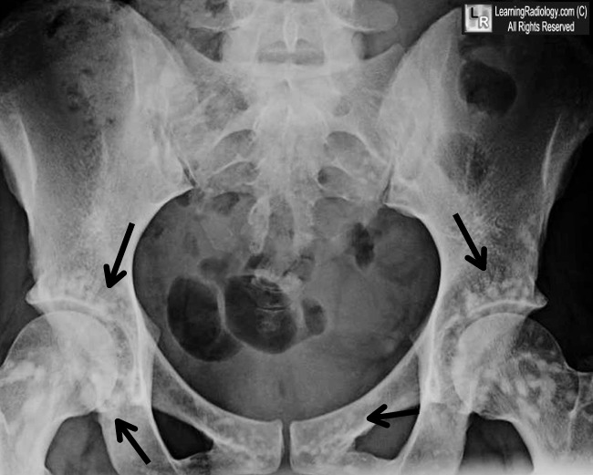
  • Well-defined sclerotic lesions clustered symmetrically around joints
  • The long axis of the lesion is typically lined-up with the long axis of the bone
  • Bone islands may have a thorny appearance
  • Low signal intensity on T1 and T2 weighted MRI images
  • Bone scan is normal
Differential Diagnosis
Treatment
  • None required
Complications
  • Associations may include connective tissue nevi called dermatofibrosis lenticularis disseminate which, along with osteopoikilosis, comprises the Buschke-Ollendorff syndrome
  • Also associated with keloid formation, dwarfism, spinal stenosis, dystocia, tuberous sclerosis and scleroderma 
osteopoikilosis
osteopoikilosis
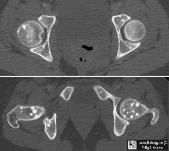
Osteopoikilosis. Black arrows point to numerous sclerotic bone islands surrounding the hip joints in a pattern characteristic of ostepoikilosis. CT images of the same patient show the well-circumscribed lesions in the femurs and pelvis.
For these same photos without the arrows, click here and here
For more information, click on the link if you see this icon 
Osteopoikilosis: A Case Report. Khot R, Sikarwar JS, Gupta RP, Sharma GL. Ind J Radiol Imag 2005 15:4:453-454

Haglund´s Syndrome


martes, 18 de febrero de 2014

Routes of migration of the femoral head


lunes, 17 de febrero de 2014

Dr Jay Smith from Mayo Clinic, Publishes New Platelet Rich Plasma Review Article: Comparing PRP Injections with Extracorporeal Shock-wave Therapy for Athletes with Chronic Patellar Tendinopathy


Julie Patchis

Dr Jay Smith from Mayo Clinic, Publishes New Platelet Rich Plasma Review Article: Comparing PRP Injections with Extracorporeal Shock-wave Therapy for Athletes with Chronic Patellar Tendinopathy

Upcoming speaker at The Orthobiologic Institute (TOBI) 5th Annual PRP & Regenerative Medicine Symposium with Cadaver Lab, Dr Jay Smith, recently published a review in the Clinical Journal of Sports Medicine of a study done by Vetrano comparing the effectiveness of Platelet Rich Plasma (PRP) versus Extracorporeal Shock-wave Therapy (ESWT) for athletes with chronic patellar tendinopathy, “jumper’s knee”.

The randomized controlled study examined 46 individuals with ultrasound diagnosed unilateral Jumper’s knee for greater than 6 months.

Twenty-three patients underwent 2 PRP injections directly into the affected patellar tendon, separated by 1 week. While, twenty- three patients underwent 3 treatments of ESWT separated by 48-72 hours. One week post-procedure, each treatment group initiated a stretching and strengthening program for 2 weeks duration, and gradually resumed normal daily and athletic activity by 4 weeks post-treatment.

The results of the 12-month follow up period showed significant improvements from baseline in both the PRP and ESWT groups, assessed via the Victorian Institute of Sports Assessment-Patella (VISA-P) questionnaire and Visual Analogue Scale during 5 single leg squats. The PRP group illustrated significantly greater improvements in VISA-P scores at 6 and 12 months follow-up, while also revealing similar improvements in VAS scores during single leg squatting. Furthermore, at the end of the 12 months follow-up period, 91% of PRP patients rated their “response to treatment” as good or excellent, in comparison to only 60.8% in the ESWT group.

The study illustrates the positive effects of both extracorporeal shockwave therapy and intra-tendon PRP injection on chronic patellar tendinopathy. However, the results of the study show greater 6 months improvements and better overall functional outcomes and patient satisfaction at 12 months with PRP treatment. Although additional studies are needed to confirm these findings and further investigate PRP efficacy, this study provides preliminary data illustrating the potential therapeutic effects of PRP in the treatment of chronic “jumper’s knee”.


Review Abstract: http://www.ncbi.nlm.nih.gov/pubmed/24366015
Study Abstract: http://www.ncbi.nlm.nih.gov/pubmed/23408591