Artroscopia Pediatrica

Artroscopia Pediatrica

sábado, 22 de febrero de 2014

Osteopoikilosis

http://www.learningradiology.com/archives2010/COW%20415-Osteopoikilosis/osteopoikcorrect.htm


Osteopoikilosis
"Spotted Bone Disease"
General Considerations
  • Rare autosomal dominant or sporadic osteosclerotic dysplasia
  • Multiple punctate sclerotic lesions representing "bone islands" or foci of compact bone located in cancellous bone
  • Asymptomatic
  • Occurs in the epiphyses and metaphyses with predilection for
    • Tubular bones of the hands and feet
    • Carpals
    • Tarsals
    • Pelvis
    • Scapula
  • Ribs, clavicles, spine, and skull are rarely involved
  • Usually clustered around joints
  • Males and females affected equally
Clinical Findings
  • Asymptomatic
  • Diagnosis is usually made incidentally
Imaging Findings
  • Well-defined sclerotic lesions clustered symmetrically around joints
  • The long axis of the lesion is typically lined-up with the long axis of the bone
  • Bone islands may have a thorny appearance
  • Low signal intensity on T1 and T2 weighted MRI images
  • Bone scan is normal
Differential Diagnosis
Treatment
  • None required
Complications
  • Associations may include connective tissue nevi called dermatofibrosis lenticularis disseminate which, along with osteopoikilosis, comprises the Buschke-Ollendorff syndrome
  • Also associated with keloid formation, dwarfism, spinal stenosis, dystocia, tuberous sclerosis and scleroderma 
osteopoikilosis
osteopoikilosis
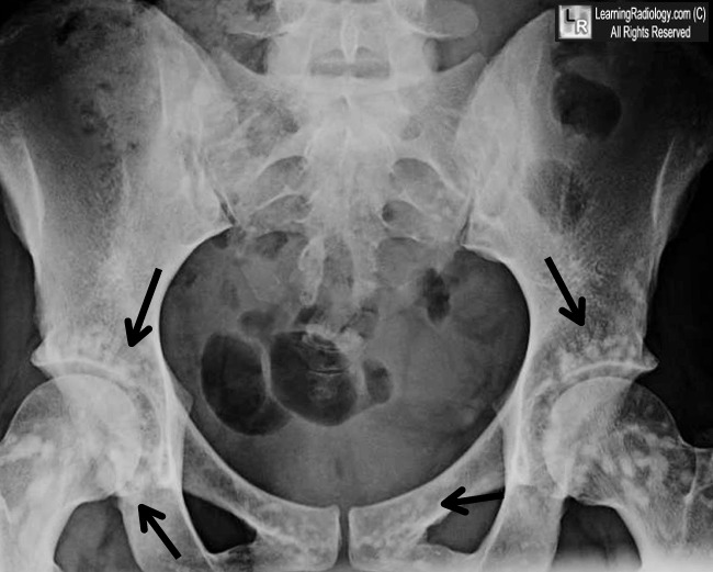
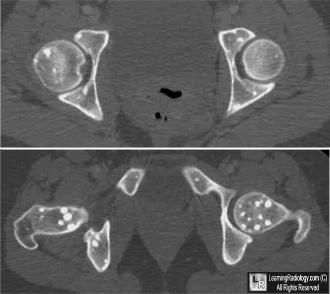
Osteopoikilosis. Black arrows point to numerous sclerotic bone islands surrounding the hip joints in a pattern characteristic of ostepoikilosis. CT images of the same patient show the well-circumscribed lesions in the femurs and pelvis.
For these same photos without the arrows, click here and here
For more information, click on the link if you see this icon 
Osteopoikilosis: A Case Report. Khot R, Sikarwar JS, Gupta RP, Sharma GL. Ind J Radiol Imag 2005 15:4:453-454

No hay comentarios:

Publicar un comentario